Log in


OASIS is the official newsletter for the Ophthalmic Anesthesia Society (OAS) and the primary source of information among specialists who treat patients undergoing cataract and other ophthalmic surgical procedures. OASIS delivers organization news, industry updates and clinical information that can be applied immediately in practices large and small. The newsletter is also a dedicated resource for anesthesiologists, ophthalmologists, certified registered nurse anesthetists and other professional personnel who are looking for niche techniques and cutting-edge research. 

To learn more about OASIS or to submit a post for the society, please contact info@eyeanesthesia.org 


  • 26 Feb 2020 1:11 PM | Anonymous

    Howard Palte, MBChB
    University of Miami System of Health 

    Ocular trauma is common arising secondary to sports injury, road accidents, assault and leisure. The quoted incidence is imprecise because estimates are retrospective and drawn from hospital records and population surveys. However, the World Health Organization (WHO) estimates there are 55 million injuries globally with more than 2.5 million occurring in the United States, alone. The majority (90%) occur in males, particularly those in the 15-24-year age group.

    The Ocular Trauma Study Group divides eye trauma into open and closed globe injury with further classification according to type, grade, pupil response and zone of injury (Table 1). Moreover, the area affected is divided into three zones, viz. zone I (limited to the cornea and limbus), zone II (< 5mm posterior to limbus) and zone III ( > 5mm posterior to limbus).

    The primary considerations in the management of eye trauma are control of intraocular pressure (IOP) and preservation of ocular integrity by preventing extrusion of ocular contents. In brief, the primary factors affecting IOP are changes in intraocular volume (blood and aqueous humor), orbit compliance and external compressive forces. Intraoperative coughing, straining and retching result in massive increases in IOP and pose significant threat to visual outcome.

    Anesthesia management of ocular trauma extends beyond the eye., The preoperative evaluation is important because there may be additional factors that modify the anesthesia plan. The provider should determine the mechanism of injury and examine for associated injuries, especially head trauma. In certain instances, additional investigations  such as, CT scan orbit and head and metabolic panel may be warranted.

    For decades, general anesthesia (GA) has been preferred for repair of open globe injuries (OGI) because it provides ocular akinesia, lowered IOP, airway control and eliminates the peril of unexpected patient movement. Anecdotal reports, dating back to the 1950s, stressed avoidance of succinylcholine because of elevations in IOP and extrusion of ocular contents. However, succinylcholine  may be considered in select OGI when airway control or aspiration is of concern because it produces moderate and transient increases in IOP (9mmHg), and not an implicated precursor of vitreous expulsion. The drawbacks of GA  include risk of regurgitation, increased nausea and vomiting and significant increases in IOP with coughing and bucking.

    Regional anesthesia (RA) with monitored anesthesia care (MAC) is an alternative option in select open globe injuries. A retrospective review at a leading eye institute examined outcomes of eye injuries managed under RA + MAC in terms of  long-term visual acuity. It found that anterior (Zone I & II) injuries and intraocular foreign bodies (IOFB) managed under RA & MAC resulted in reduced operative times, and no statistical difference in visual acuity at 2-month follow-up.

    The regional anesthesia technique used in open globe injuries requires modification in order to prevent fluctuations in IOP. Eyelid squeezing causes massive increases in IOP and is attenuated by performance of a facial nerve (VII) block, commonly adopting a modified van Lint approach. The subsequent extraconal (peribulbar) block is performed using a restricted volume (5-6 ml) of LA injected in small aliquots over 2-3 minutes (limiting external compressive forces on the globe).

    Eye injuries in children require operative repair under GA. It is estimated that 90% of these injuries are preventable because the majority occur at home when there is no parental supervision.  The mechanism varies with age with cleaning products injuring toddlers and older children more likely to sustain sports and projectile insults.

    Primary closure of open globe injuries is optimally achieved within 24 hours. Long-term visual outcomes are determined by initial visual acuity . The US Eye Injury Register indicates that long-term visual acuity is determined by presenting light perception. Ultimately, 3 /4 patients retain a degree of vision and 20% regain normal light perception. 


  • 12 Nov 2019 12:09 PM | Anonymous

    First off, I would like to sincerely thank the Board of Directors and all the members of the Ophthalmic Anesthesia Society for your trust and support in nominating me as this year’s president. I will do my best to fulfill my duties and responsibilities, but I will need your help - I want to hear your ideas, feedback, and suggestions to help our society develop a strong voice for patient care, patient safety, and operating room business in ophthalmic surgery and anesthesia.

    Membership is very crucial for the survival of any organization, and I believe that the value of the membership is a very important factor in retaining existing members and attracting new members. Therefore, improving the value of our membership will continue to be my main focus of the OAS business in the upcoming year.

    The OAS annual scientific meeting has been a great platform for all our members to come together face to face to exchange updated information, discuss new founded theories, and practice ophthalmic surgery and anesthesia. I want to enhance the educational value of the 34th Annual Scientific Meeting and start the planning of the 34th annual scientific meeting early in order to invite valuable speakers, to advertise the benefits of the meeting, and attract new members to our society while gaining new attendees.

    Many of our members are already experts in the practice of ophthalmic surgery and anesthesia. Their publications, including papers, articles, abstracts, practice protocols, institutional policies, and letters to editors are very valuable to showcase to our members and the international ophthalmic society as well. Four years ago, during my first term of presidency, I wanted to create an online repository to host all the publications from our members, however, the project was delayed due to technical difficulty so I would like to try to implement this one again in the upcoming year. In addition to post your work, the repository will also house guidelines from centers across the country to help in building consensus statements on key issues in the specialty. I will need each of you to send all of your publications to Lori and Melissa as the first step of this exciting project.

    The OAS newsletter has been getting better and better in the last several years thanks to the hard work of the OAS Scientific Advisory Panel, but we need help from every member to participate and make a contribution to the newsletter to make it more meaningful and educational. Please send your suggestions to Lori, Melissa, the board members, and myself included.

    The online discussion forum has also been a great success in the last several years by being more simple, effective, and accessible. If you have questions or have answers to the questions other members posted, we encourage you to participate as this is can be a great learning experience for you and your peers. Our small society is advantageous for us to get to know each other, so I hope the improvement of the online discussion forum can further develop the communication between our members.

    In addition to improving the value of membership, I am also asking every member to help our society grow by reaching out to our surgical and anesthesia colleagues and encouraging them to become new members and participate in this wonderful community that we have built.

    If you have any questions, comments, or concerns, please don’t hesitate to reach out to me. I am very excited for the new business year of the Ophthalmic Anesthesia Society and am looking forward to working with every member closely for the bright future of our society.


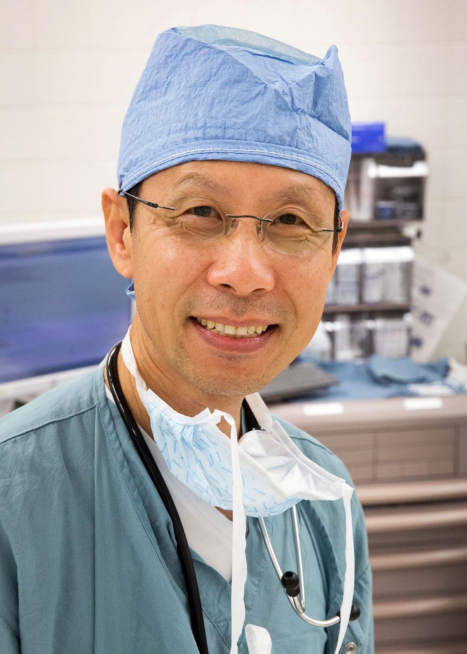
    Respectfully submitted, 

    Zhuang T. Fang, M.D., MSPH, FASA
    Clinical Professor
    David Geffen School of Medicine at UCLA
    Department of Anesthesiology and Perioperative Medicine
    Associate Director, Wasserman Stein Operating Service


  • 12 Nov 2019 11:49 AM | Anonymous

    This fall, TLC presented data at the American Society of Anesthesiologists annual meeting from a Phase I/II clinical trial which showed TLC590 to yield more immediate and long-lasting pain reduction than ropivacaine.

    The multilamellar vesicles ropivacaine (MVR) demonstrates a sustained-release profile in an in vitro serum environment model after 24 hrs of incubation which translates in the in vivo rat pharmacokinetic profile of ropivacaine as a prolonged half-life that is 10-fold longer in duration than plain ropivacaine solution. The anesthetic effect of single-dose MVR is apparent by providing a prolonged analgesia effect compared to plain ropivacaine solution in an in vivo guinea pig pin-prick wheal model after a single intracutaneous injection.

    From a safety evaluation, MVR is well tolerated after a subcutaneously injection at a dose level of 20 mg/kg in rats, with no observable changes in clinical observation, body weight, organ weight, hematology and serum chemistry analysis In addition, in vivo findings in which TLC590 showed no dose-related toxicity and other preclinical data were recently published in the International Journal of Nanomedicine.

    OAS members have long expressed interest in non-opioid solutions for pain management following surgery, so we’ll stay tuned for more advances – including advances in nanoscience and bioengineering.

    Read the journal article online.

  • 12 Nov 2019 11:49 AM | Anonymous

    This month, the Centers for Medicare and Medicaid Services (CMS) has released the 2020 Medicare Physician Fee Schedule final rule, which includes the proposed drop in payments for cataract surgery.

    The final work relative value unit (RVU) for complex cataracts 66982 is 10.25, compared to the current Work RVU of 11.08, a $47 reduction. For non-complicated cataracts 66984, the proposed Work RVU is 7.35, compared to the current 8.52, a $97 reduction.

    Improved technology in hands of a skilled ophthalmic surgery care team have created efficiencies and, ultimately, a reduction in time. The decrease in valuation is directly connected to the decreasing time ophthalmologists spend in the surgery room and the decreased time spent on post-operative visits (especially the less frequent, fourth post-operative visit).

    The American Academy of Ophthalmology (AAO) and the American Society of Cataract and Refractive Surgery (ASCRS), meanwhile, have lobbied for a less dramatic decrease in reimbursements by highlighting on the other part of the RVU’s formula: intensity and complexity.

    Cataract surgery is a complex procedure with irrefutable, life-changing outcomes.

    So, what advice are the experts giving to blunt the decrease in Medicare/Medicaid reimbursements?

    • Ophthalmic surgery teams should pay their dues and become more active with their professional societies. Ongoing collaboration with OAS, AAO, ASCRS, and the American Society of Ophthalmic Administrators will empower these organizations to ensure we are represented whenever RVUs are considered. RVUs are determined by a special committee formed by the American Medical Association (AMA), known as the AMA/Specialty Society RVS Update Committee. We need seats at their table!
    • Reevaluate your cases. It may be prudent to audit charts more judiciously, confirm that visual criteria are clearly defined and each patient truly needs cataract surgery.
    • Streamline services and diversify, where appropriate. Some patients may benefit by undergoing other procedures in conjunction with cataract surgery: glaucoma techniques, femto-LASIK surgery, multifocal lens implants, etc.  
    • Continue developing relationships with your patients and delivering quality care. The reason ophthalmic surgery procedures weren’t cut as dramatically as we feared is because there is tremendous value to the care we provide. Our skills, the quality of our care and our outcomes are a factor when considering the intensity and complexity in valuation discussions.


    Learn more:

    https://www.federalregister.gov/documents/2019/11/15/2019-24086/medicare-program-cy-2020-revisions-to-payment-policies-under-the-physician-fee-schedule-and-other

    http://ascrs.org/2020-medicare-physician-fee-schedule-mpfs-proposed-rule

    https://www.aao.org/eyenet/article/cataract-surgery-payment-in-medicare?september-2019

    https://asoa.org/news/ascrs-special-report-key-information-about-2020-medicare-physician-fee-schedule-proposed-rule

    https://www.reviewofophthalmology.com/article/medicare-proposes-another-cut-in-cataract-reimbursement


  • 14 Aug 2019 1:48 PM | Anonymous
    By Ric Rivers, MD, and Tina Tran, MD

    OAS is happy to roll out a new series, designed to connect members from across the United States and showcase our communities. Ophthalmic anesthesia is a very niche field so we're working hard to give member every opportunity to connect, discuss and discover. 

    The first in the series spotlights the Wilmer Eye Institute. We hope you enjoy it! 


    This summer marks the 10-year anniversary of the opening of Wilmer Eye Institute’s Robert H. & Clarice Smith Building and Maurice Bendann Surgical Pavilion.

    The first of its kind, this landmark building united ophthalmology research and care delivery to a degree not seen before in the field of ophthalmology. The building supports engineers, scientists, clinicians and their teams in working together and sharing ideas in an open, interactive, and collaborative environment. 

    In light of this anniversary, here is a quick introduction to the Wilmer Eye Institute written by OAS members Ric Rivers, MD, and Tina Tran, MD.

    About William Holland Wilmer & Wilmer Eye Institute 

    William Holland Wilmer began to practice ophthalmology in 1889 and over the next 35 years became one of the most renowned practitioners in his field. In 1924, the Wilmer Foundation was started to raise funds to allow him to carry on his own research work and to train young doctors. An endowment of nearly $4 million was raised, and one year later, the foundation established the Wilmer Institute of Ophthalmology at The Johns Hopkins Hospital. The institute was the nation's first university eye clinic to combine patient care, research and teaching under one roof, and Wilmer was chosen, at age 61, to become its inaugural director. He remained in that position until his retirement in 1934.

    At the beginning there was an ophthalmology emergency room, two ophthalmology operating rooms, and eight ophthalmology inpatient beds in a single same Wilmer building, spread over 2 floors. About 15 years ago we finally grew out of that space. Now Wilmer shares space in the Johns Hopkins adult emergency department, has seven ophthalmology operating rooms in the new Smith building, and admits patients to the main hospital. Wilmer currently performs almost 7000 cases per year and all subspecialties are represented.

    The operating room operates with a team model that relies a couple of monthly meetings where nursing, surgery, anesthesia, materials management, and administration all participate to make an efficient operation. Patient care is optimized through the use of an electronic medical record that is shared by all members of the team. Daily routine includes morning debriefing sessions where nursing and anesthesia discuss the issues of the day whether it is related to equipment, personnel, or patients.

    The seven dedicated operating rooms are separated from the main hospital in a separate building, across the street. All ophthalmology surgical cases except the smallest peds patients, and the critically ill, are performed in this facility. Ninety-five percent of the patients are outpatients and about five percent are planned post-op admissions (transferred by hospital-owned ambulance). 

  • 07 Aug 2019 10:52 AM | Anonymous

    A recent study, published by MBJ Open Ophthalmology, set out to determine if automated OCT aids in the management of patients in a high-volume after-hours clinic.

    OCT has become the standard of care for diagnosing plenty of ocular pathologies, but is generally not available at most institutions after-hours -- when many patients present with acute complaints. Richard I. Kaplan, MD, New York Eye and Ear Infirmary of Mount Sinai, and his team, obtained images over a period of 15 months (202 patients, 359 eyes), reviewed electronic patient records, and surveyed staff to measure the advantages of having an automated OCT for diagnosis and management of acute complaints and ocular emergencies.

    Chief complaint, image type, image findings and diagnosis were analyzed. Ultimately, patient complaints included flashes/floaters, metamorphopsia, decreased vision and scotomas. Diagnoses comprised of vascular occlusion, retinal detachment, macular hole, cystoid macular edema and central serous retinopathy.

    Twenty-five residents and fellows participated in the voluntary survey. All residents (n=18) and fellows (n=7) found the OCT to be helpful; 15 residents (83%) and 4 fellows (57%) felt that it changed management. Most participants, (21 or 83%), felt that the OCT improved patient satisfaction and reduced delayed or misdiagnosis. Most participants (19 or 76%) also reported reduced physician stress.

    Kaplan et al noted OCT aids in the quickly-needed diagnosis of central retinal artery occlusion (CRAO) – data showed six patients with CRAO, and OCT successfully confirmed five.

    They also highlighted OCT’s use in differentiating macular-involving versus macular-sparing retinal detachment, leading urgent retinal detachment repair or surgical planning and patient couselling. “In cases of macula-off retinal detachments, the OCT provides objective documentation. As most ophthalmic lawsuits in the USA relate to RD, such documentation substantiates the appropriateness of care in the after-hours setting,” Kaplan explained.

    OCT was also found helpful in ruling-out emergent pathology, thus refining triage of patients to subspecialty clinics in hospital and reducing the need for after-hours subspecialty consults. The study included 14 patients with central serous retinopathy who, in general, with OCT verification, follow-up appointments could be extended to 3 weeks.

    Although trauma cases were not well represented in the sample, OCT appears to aid in the identification of globe perforation and may aid in ruling out such injuries in the emergency setting.

    Finally, one limitation of automatic OCT is the minimum vision requirement necessary for patients to find fixation cues without operator redirection. Some attempted scans had insufficient signal strength, all in patients with visual acuity 20/400 or worse.

    “In the after-hours or acute-care setting, the most important goal of care is the timely diagnosis and treatment of vision threatening pathology while efficiently using ancillary testing and consultants. We found OCT accessibility in the after-hours setting improved the diagnosis and disposition of patients. Additional studies will be helpful to further appreciate the benefits of including OCT alongside clinical examination in both eye-only and general emergency settings.”


  • 02 Aug 2019 10:56 AM | Anonymous

    Summer greetings! Its late July and our 33rd Annual Scientific Meeting (September 13-15, 2019) is almost upon us.

    I am excited at the variety and depth of topics to be covered and am confident that the program will offer special value to attendees whether they are anesthesiologists, ophthalmologists, CRNAs or allied professionals. We are indeed fortunate to host top local experts as well as a number of esteemed international guests. Their combined experience has not been paralleled at prior meetings. In fact, this assembly will offer attendees a rare opportunity to glean valuable theoretic and practical knowledge in a condensed 2-day span.

    Many of our local faculty is well known to you. They offer attendees the benefit of invaluable knowledge and years of experience gained from practice at their academic institution or private practice. I am confident that each lecture will leave you with at least one ‘pearl of wisdom’.

    In the interest of brevity, I want to whet your appetites with a peep at a few of our faculty. Steve Gayer, MD, MBA, is an internationally acclaimed, widely published authority on ophthalmic anesthesia. Similarly, we are fortunate to host Chandra Kumar, MB BS a well-respected academician and keen proponent of sub-Tenon anesthesia. And to complement them we will benefit from the erudite and articulate deliveries of British ophthalmologist, Tom Eke, MA (Cantab), MD, FRCOphth. In addition, we host Sujatha Ravichandran, DA, DNB, a pediatric anesthesiologist from India. The eye block workshop is not to be missed because brief technique presentations are complemented by hands-on exposure in cadaver and porcine eye models. And, world-class experts in needle and cannula-based regional ophthalmic anesthesia will tutor this year’s workshop!

    Finally, our program offers a great balance between didactic lectures, practical workshops, interactive group discussions and most importantly, social discourse. As a reminder, registration is now open on our website (eyeanesthesia.org) and early-bird registration extends through August 24.

    OAS Chicago 2019 is one meeting you don’t want to miss! I look forward to welcoming you.

    Warm regards,
    Howard Palte, MBChB, FCA Bascom Palmer Eye Institute


  • 14 May 2019 12:55 PM | Anonymous

    Greetings!

    September is fast approaching and the organizing committee is now making final adjustments to the great program for our 33rd Annual Scientific Meeting. We look forward to seeing as many of you as possible in Chicago, and earnestly urge you to encourage non-member colleagues and associates to join us! 

    We have assembled a prestigious, diverse group of speakers, many of whom have already confirmed their participation. So, I am excited to introduce some faculty who are traveling from abroad: 

    • Chandra M Kumar, MBBS, senior consultant in anesthesia for Khoo Teck Puat Hospital (Singapore) and a Professor of Practice/Visiting Professor for Newcastle University & Newcastle University Medical School (UK). Chandra is a widely-respected author of many ophthalmic anesthesia papers.
    • Tom Eke, MA, MD, FRCOphth, consultant ophthalmologist for Norfolk and Norwich University Hospitals (UK). Tom’s main research interests include the safety and effectiveness of local anesthesia, and patient-centered outcomes in glaucoma surgery.
    • Sujatha Ravichandran, DA, DNB, who practices at the not-for-profit charitable eye hospital, Sankara Nethralaya, in India. 

    I would be remiss not to remind you that registration is now open and we are also currently accepting abstract submissions. This is a unique opportunity for members from all disciplines to present case studies, research data or technique modifications to an audience of your peers – so, please get to it and submit your abstracts ahead of the August deadline.

    I am also extending an open invitation to your local network members to join our society. As an incentive, new members will receive a 15% discount on registration. When you enroll online at the OAS website (eyeanethesia.org), the new member activation email will include a code that automatically applies the reduced registration fee. 

    I am always available via email (hpalte@miami.edu) to accept ideas or address inquiries, so don’t remain a stranger!

    Howard Palte, MBchB
    OAS President


  • 14 May 2019 12:05 PM | Anonymous

    Another clinical study published in Ophthalmology, this one led by Crandall Peeler, MD, of Boston Medical Center, examined oral sedation versus intravenous sedation. 85 patients were randomized to receive either oral triazolam with IV placebo or IV midazolam with oral placebo preoperatively.

    The primary outcome was patient satisfaction, measured by a survey administered on postoperative day one. Secondary outcomes included surgeon and anesthesia provider satisfaction, need for supplemental anesthesia, and surgical complications.

    Mean patient satisfaction score was 5.34 ± 0.63 (range 3.75 to 6) in the oral sedation group and 5.40 ± 0.52 (range 4 to 6) in the IV group. Eight patients in the oral group (19.0%) and 3 in the IV group (7.0%) received supplemental IV sedation. Read the full study here.


  • 14 May 2019 11:33 AM | Anonymous

    By Maggie Jeffries, MD

    Peribulbar blocks have similar success rates to retrobulbar blocks. Due to the absence of an intermuscular membrane to separate extra- from intraconal compartments, peribulbar injections result in a similar space for spread of local anesthetics. During a peribulbar block, local anesthetic spreads into the adipose tissue of the orbit, including the intraconal space where the nerves (motor & sensory) to be blocked are located.  Unique to peribulbar and NOT retrobulbar injections, local anesthetic spreads to the lids to block the orbicularis muscle and often obviates need for supplemental lid block.  Lid squeezing is an undesirable motor action that is not blocked by a retrobulbar injection.1

    Because peribulbar blocks are extraconal injections, there is less risk of complications such as optic nerve injury, brainstem anesthesia, and retrobulbar hemorrhage. Caution must still be taken as myopic staphyloma, which occur in highly myopic eyes (“long”, >26mm), could lead to globe perforation. As with all blocks, local anesthetic spread can be uncertain or incomplete necessitating another block to be performed.1

    Peribulbar blocks are typically performed using a 25 gauge 1” sharp or Atkinson needle. The needle is inserted at the inferotemporal corner of the eye at the junction of the lateral 1/3 and medial 2/3 of the lower orbital rim. The needle is passed posteriorly, parallel to the floor of the orbit until it is estimated to lie beyond the equator of the globe. A volume of 5–10 ml of local anesthetic is injected after negative aspiration.2


    #2 Medial caruncle

    #4 Insertion of needle for a peribulbar block.3

    A medial canthal block is a great supplement to an infero-temporal peribulbar block when complete akinesia is desired (e.g. corneal transplant, retina).  This blocks the medial rectus muscle which is a muscle often missed with a standard peribulbar block.  A superior nasal block will also block the medial rectus and superior oblique but is a riskier block due to location in relation to orbit (risk for perforation) and vascular supply.  The medial canthus is an avascular location and lacks vital anatomic structures.

    A medial canthal block is performed using a 27-gauge ½” needle.  The needle is inserted medially to the caruncle at the medial end of the lid aperture, aiming towards the nose at about a 30-degree angle. Approximately 2ml of local anesthetic is injected and you can often feel it spreading around globe with the fingers holding the lids open.  Some bleeding at the medial canthus can be expected but it is usually minimal and self-limited.  Caution should be taken as this block can induce sneezing so be prepared if patient has sharp inhale.  When using the shorter needle, ones doesn’t need to worry about needle depth.1,4

    Facial nerve blocks are more commonly needed with retrobulbar blocks as there isn’t spread through the orbital fat to the orbicularis muscle but can be used with any block.  This block is particularly useful in the setting of scleral buckle retina procedure where lid manipulation often triggers patient discomfort.  A facial nerve block is also great for patients who squint against the lid speculum.  The Van Lindt facial nerve block is the most common performed in ophthalmology and involves injecting local anesthetic at the crossing between a vertical line 1 cm lateral of the outer orbital rim and a horizontal line 1 cm below the inferior orbital rim.6 An additional benefit of the use of a facial nerve block is that the facial nerve innervates the conjunctiva and thus blocking this nerve provides additional conjunctival anesthesia.7

    Summary:

    Peribulbar and medial canthal blocks, when used separately or together, can be just as effective retrobulbar blocks. They can provide complete akinesia and analgesia when performed properly and, with the addition of a facial nerve block, can provide additional akinesia and analgesia of the eyelid. The facial nerve block will also provide some sensory block of the conjunctiva.

    References

    1. Ripart J, Mehrige K, Della Rocca, R. Local & Regional Anesthesia for Eye Surgery. NYSORA https://www.nysora.com/local-regional-anesthesia-for-eye-surgery
    2. Lopatka CW, Magnante DO, Sharvelle DJ, Kowalski PV. Ophthalmic blocks at the medial canthus. Anesthesiology 2001;95:1533.
    3. Ripart J, Lefrant JY, Vivien B,  Charavel P, Fabbro-Peray P. Ophthalmic Regional Anesthesia: Medial Canthus Episcleral (Sub-Tenon) Anesthesia Is More Efficient than Peribulbar Anesthesia: A Double-blind Randomized Study. Anesthesiology 2000; 92: 1278-1285.
    4. Hustead RF, Hamilton RC, Loken RG. Periocular local anesthesia: medial orbital as an alternative to superior nasal injection. J Cataract Refract Surg 1994 Mar; 20(2):197-201.
    5. Anker R, Kaur N. Regional anaesthesia for ophthalmic surgery. BJA Education 2017 July; 17(7): 221–227.
    6. Schimek F, Fahle M. Techniques of facial nerve block. British Journal of Ophthalmology 1995;79:166-173.
    7. Ruskell GL. Innervation of the conjunctiva. Trans Ophthalmol Soc UK 1985; 104(pt 4): 390-5.


Copyright © 2016 Ophthalmic Anesthesia Society. All Rights Reserved.

Powered by Wild Apricot Membership Software